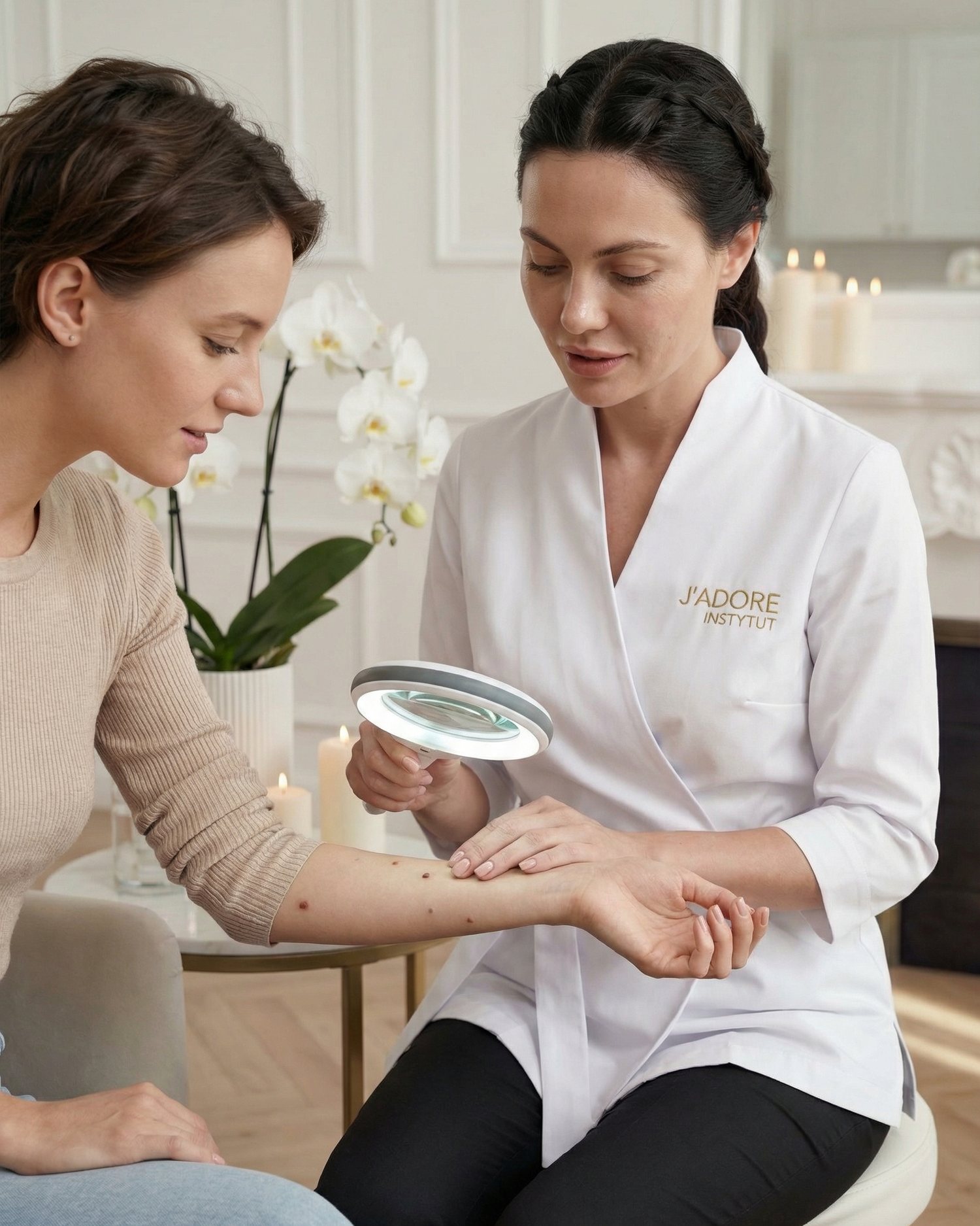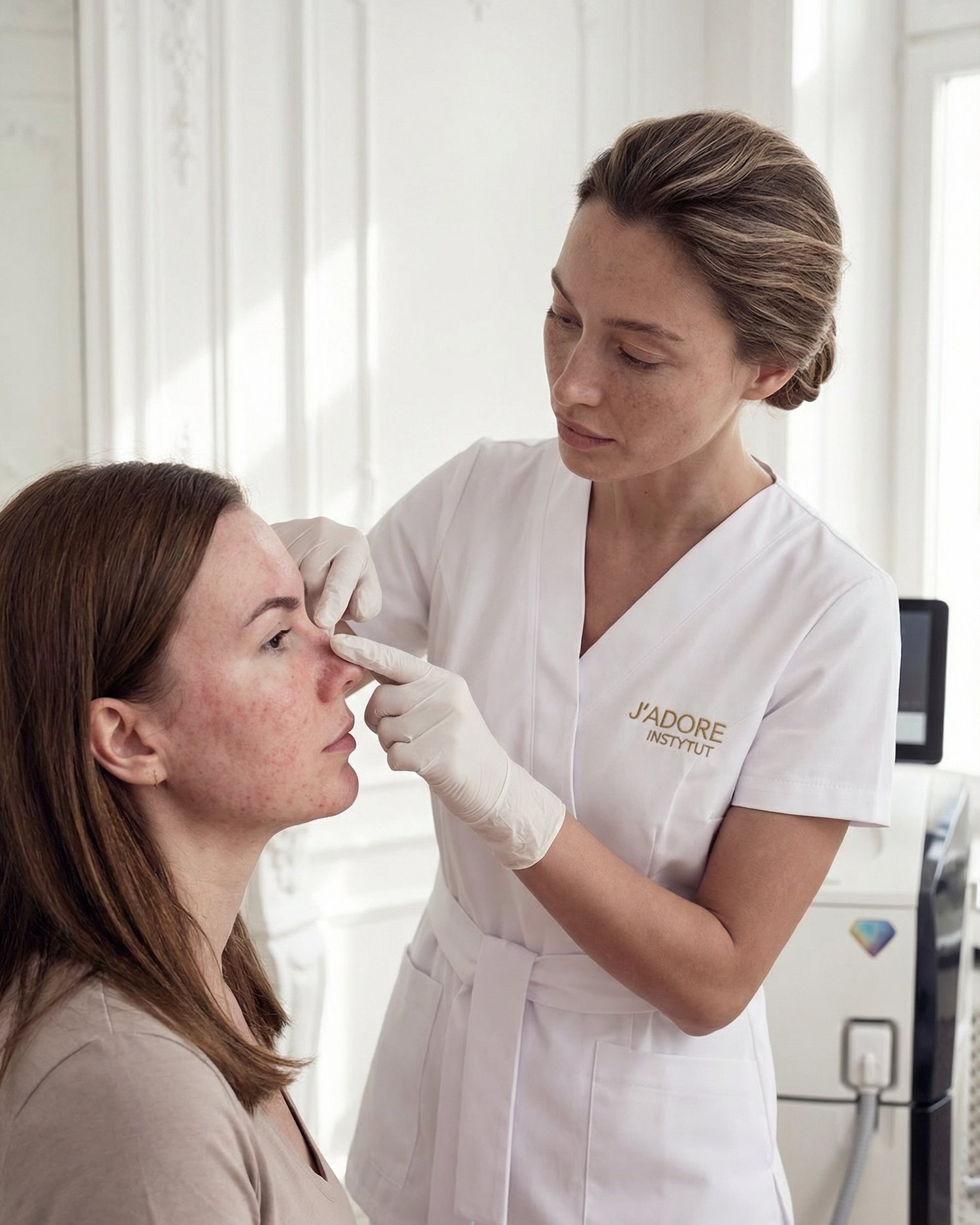
The first criterion is: does the tissue need to be sent for examination?
Electrocoagulation, cryotherapy and some laser treatments destroy the lesion on the spot. Excision, punch biopsy or shave biopsy can preserve material for microscopic assessment, depending on the technique. When there is a suspicion of cancer or the diagnosis is uncertain, the possibility of histopathology carries more weight than the speed of the procedure.
The American Academy of Dermatology indicates that biopsy is the way to confirm many skin diseases, including cancers. Electrocoagulation is therefore not a "less invasive biopsy". It is a destructive procedure. If the doctor needs the architecture of the entire lesion and its margins, thermal removal may be an inappropriate start.
Electrocoagulation works well for small, diagnosed superficial lesions.
High-frequency current generates local heat and coagulates the tissue. A thin electrode allows for targeted action, which is why the method is used for selected cherry angiomas, fibromas or seborrhoeic keratoses after qualification. The advantage is often the short working time for several small points and the ability to precisely stop bleeding.
Thermal injury remains a limitation. Action that is too deep or too wide can increase the risk of scarring, hypopigmentation or hyperpigmentation. A scab usually forms after the treatment and must fall off on its own. The method does not become traceless just because the electrode is thin.
Cryotherapy freezes, but the depth is harder to see at the time of treatment.
Cryotherapy uses a very low temperature, most often liquid nitrogen, to destroy selected tissue. It is used for various lesions, including some viral warts and keratoses, depending on the diagnosis. There is no contact with electric current, which may be relevant for some implantable devices, but it does not remove the need for a full medical history.
Laser is the name of a family of tools, not a single alternative.
- The question "laser or current" is incomplete. Lasers differ in wavelength, target in the tissue and the way energy is delivered.
- Vascular laser. It chooses haemoglobin as a chromophore.
- Ablative device and other systems. An ablative device evaporates water in the tissue, and other systems have completely different indications. The word "laser" itself does not say whether the method fits a cherry angioma, a wart or a pigmented lesion.
The advantage can be selectivity for a specific target, such as a vessel, and the ability to work without mechanical cutting. Limitations include the cost of the device, the need for proper parameter selection, eye protection and the still-present risk of burns, scarring or pigment changes. Technology does not replace diagnosis.
Excision provides material and margin control at the cost of stitches or a longer wound.
The biological price is different: the wound may require stitches, monitoring, removal of threads and limiting the stretching of the area. The scar is planned along the skin line, but it does not disappear by magic. On the face, eyelid or in an area of high tension, the choice of operator and closure technique is of particular importance.
Shave and curettage occupy a place between destruction and classic excision.
- Shave. It consists of a shallow removal of the protruding part of the lesion.
- Curettage. It consists of mechanical removal of tissue with a curette.
- Combining methods. They are often combined with a method that stops bleeding, such as electrocoagulation.
- Material for examination. In selected cases, the obtained material can be examined, although the technique and purpose of the procedure must be determined before starting.
These methods are not simply a "gentler excision". They do not always cover the full depth, and the risk of recurrence depends on the type of lesion. If melanoma is suspected, a diagnostic path determined by a dermatologist is needed, not cosmetic flattening of the surface.
The type of lesion narrows the choice more than its size.
Vascular lesion
e.g. a correctly diagnosed cherry angioma
- selective vessel closure is what counts
- electrocoagulation or an appropriate laser is considered
- colour and depth influence the choice of energy
Lesion requiring diagnosis
new, changing or ambiguous
- dermoscopy has priority
- material for histopathology is often needed
- a destructive method may be inappropriate
A soft fibroma on a stalk, a flat seborrhoeic keratosis, a viral wart and a melanocytic naevus may have a similar size, but a different structure and different risks. A ranking of devices makes no sense without naming the tissue they are to work on.
The face, eyelid, neck and sole heal according to different rules.
On the face, precision and protection of anatomical structures count. On the eyelid, there is also the proximity of the eye, thin skin and swelling. The neck and décolletage are exposed to the sun and the friction of jewellery. The sole bears the body's weight, so even a small wound can make walking difficult. The same method does not have an identical balance in every location.
The operator takes into account the possibility of a dressing, skin tension, visibility of the future scar and the risk of pigmentation. The duration of the treatment is often the least important parameter. A few minutes saved in the clinic do not compensate for weeks of poorly planned healing in a place constantly rubbed by a strap or shoe.
Phototype and a tendency to hyperpigmentation change the energy balance.
Every controlled injury can trigger post-inflammatory hyperpigmentation. The risk depends on the phototype, inflammation, depth of damage, sun and individual reaction. A laser is not automatically safer for darker skin; you must choose the right system and parameters. Cryotherapy can leave a lighter mark if it damages melanocytes.
Electrocoagulation can also end in a spot darker or lighter than the surroundings. Before deciding, show previous scars and tell us how bites, acne or cuts healed. The history of your skin is more practical information than the "no marks" promise on a device's website.
A pacemaker does not choose cryotherapy for you, but it changes the qualification for current.
With an implanted pacemaker or defibrillator, electrosurgery requires a special procedure and sometimes a cardiology consultation. The type of current, its path of flow and distance from the device matter. You cannot draw a universal conclusion based on the name of the method alone.
Cryotherapy does not use the flow of current through the patient, but it may still be inappropriate for other reasons, such as circulatory disorders in the treated area. Excision, in turn, requires an assessment of clotting and medications. Every alternative shifts the focus of risk; none removes the need for a health history.
A viral wart shows why one session does not always close the topic.
A wart is not just excess tissue. Its connection with the HPV virus and local immune response means that it can recur or require a series. Cryotherapy, curettage, electrosurgery and topical preparations have different applications depending on the number, location, duration and age of the patient.
Destruction of the visible part does not guarantee that the problem will not return. On the sole, one must additionally distinguish a wart from a corn and plan the loading of the foot. The promise of "one visit and it's over" is too simple for a lesion whose behaviour depends on more than just the depth of the device.
A cherry angioma rewards precision, but diagnosis still precedes the impulse.
A cherry angioma is a benign vascular lesion. Small points on the torso can be considered for electrocoagulation or a vascular laser. The choice depends on the number, diameter, colour, available device and the operator's experience. With many lesions, the total area of healing also matters, not just the time of each impulse.
The red colour is not a diagnosis in itself. A lesion that bleeds, grows quickly or looks different from the others requires re-evaluation. Even a typical cherry angioma can be removed correctly only when the person qualifying is sure that it really is one.
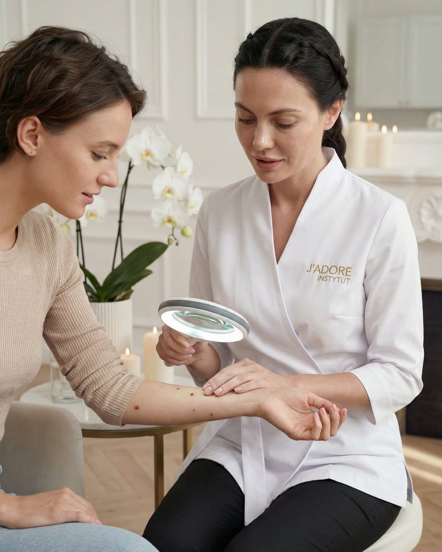
A fibroma on the neck and a mole on the neck are two different conversations.
A soft fibroma often hangs on a narrow stalk and is irritated by a collar. It can be removed using various techniques, including scissors, electrosurgically or by cryotherapy, depending on the appearance. A melanocytic naevus, on the other hand, contains pigment information, the assessment of which may require dermoscopy and histopathology.
A patient sees "two bumps". A specialist sees different cellular origins and a different diagnostic question. This is the best example of why a price list based on the number of points should not replace qualification based on the type of lesion.
Recovery should be tailored to your life, not hidden in the fine print.
After electrocoagulation or laser treatment, a scab and redness may remain. Cryotherapy more often creates a blister and weeping. Excision leaves a wound with stitches and sometimes limits sports and stretching in the area. A shave excision heals from the surface and requires systematic dressing. These differences should be part of the decision before the treatment.
If you work in front of a camera, swim or travel to the sun, the method with the seemingly shortest procedure may have the worst timing. Ask how the wound will look the next day, after a week and after a month, and when you are allowed to return to makeup, swimming pools and UV exposure.
The scope of the first session can test the plan without removing everything.
At the next visit, documentation counts: how long the scab lasted, whether a spot formed, whether the point closed according to the plan and whether the aftercare was feasible. If the skin's response was difficult, you can change the technique, parameters, timing or abandon further removal. If the process was calm, you still must not automatically treat a dark nodule next to it as another cherry angioma. A good reaction to one method confirms the tolerance of a specific procedure in a specific place; it does not diagnose all remaining lesions.
A person returning after a fifth successful treatment may know the smell, the sensation of the pulse and the aftercare method, but each new lesion undergoes qualification again. Conversely, someone coming for the first time does not have to agree to a large scope just because many points look similar. Ask if you can start with a smaller number, when the follow-up will take place and on what basis the decision to continue will be made. This approach does not decide in advance that laser or electrocoagulation is better. It shows, however, how to move the comparison of technologies from a marketing brochure to the behaviour of your own skin.
A scar after excision may be a better result than no diagnosis.
In aesthetic conversations, a scar is often treated as a failure. However, with a suspicious lesion, a planned line after excision is the price for the material, margin and a reliable answer. Attempting to avoid a stitch at all costs can lead to the destruction of the surface without certainty about what was deeper.
On the other hand, a classic excision of a benign, tiny cherry angioma would usually be disproportionate. The right choice does not minimise the scar in isolation from the goal. It minimises the entire risk: diagnostic, functional and aesthetic.
The price should not reward a method that does not fit the lesion.
A cheaper pulse is not a saving if the lesion recurs, the material disappears without examination or a mark requiring correction is created. A more expensive laser is also not better just because the device looks modern. You pay for the qualification, the right tool, hygiene, energy control, healing instructions and access to help after the treatment.
Do not choose a salon solely based on the number of lesions included in one service. Ten correctly identified cherry angiomas and ten different, unclear nodules do not create the same task. A fair quote can only be provided after examining the skin.
Five questions filter out technological marketing.
- What is the diagnosis and who made it?
- Will material for histopathology remain after this method, and if not, why is it not needed?
- What alternatives fit this location and skin phototype?
- What will the wound look like and what is the risk of recurrence, scarring and pigment change?
- What will we do if the test result or healing does not go according to plan?
The answer "it is the best machine" is not enough. A good consultation explains the mechanism of choice without making the device name the final argument. It should also allow for a referral to a dermatologist, surgeon or another facility when the required method is not available on-site.
Sometimes the best option is observation.
A benign lesion that does not hurt, bleed, change or bother you does not always require removal. A dermatologist may recommend photographic documentation and follow-up. Every procedure creates a wound and has a risk, however small. No action is a legitimate decision if the diagnosis and observation plan are clear.
The patient's age changes the threshold of caution.
- Cooperation and consent. In a child or teenager, not only the diagnosis is important, but also cooperation during the procedure, anaesthesia and the necessity of a guardian's consent.
- Growing skin and scarring. The impact of a scar on growing skin is significant.
- Order of procedure. A lesion that looks like a wart may first be covered by conservative treatment. Rapid destruction is not always the first choice.
- Factors requiring attention. In an older person, many benign lesions are encountered more often, but the importance of anticoagulants, skin fragility and a history of cancer also increases.
- Assessment of each lesion. You should not remove an entire map of points according to one scheme. Each suspicious lesion must be separated from the typical ones.
- Scope of the session. The scope of a single session should be adjusted to the ability to care for several wounds.
At any age, the reason for the treatment is important. A child scratching a painful lesion, an adult catching a fibroma with a collar and a person wanting to remove a stable point solely for aesthetic reasons have different benefits and risk tolerance. The technique appears only after this conversation.
Age should not act as an automatic consent or prohibition. It is one of the elements alongside the diagnosis, medications, fitness, location and expectations. The same person may have a cherry angioma suitable for short electrocoagulation and another mole requiring excision. The assessment takes place lesion by lesion, never for the whole body according to one shortcut.
A consultation in Warsaw should end with a justification.
When browsing skin lesion removal in Warsaw, treat the description of electrocoagulation as one possibility, not an automatic verdict. Ask for the name of the diagnosis, the reason for the choice and the healing instructions. If an excision or examination is needed, the right answer may lead beyond this treatment.
The method is chosen only after setting a hierarchy of goals.
The hierarchy is simple: do not miss a disease, preserve the necessary material, remove the lesion effectively, protect the function of the area, and only then minimise the mark and time. When this order is reversed, aesthetics can overshadow diagnostics. When you stick to it, different technologies stop competing like car brands.
Electrocoagulation is accurate for some small, benign lesions. Cryotherapy responds well to other indications. A laser may be the most selective for a specific chromophore. Excision wins where depth and histopathology are needed. Sometimes observation wins. A conscious choice does not consist of finding one method for everything, but of accepting that the skin poses different questions.