Where the belief that a visual check is enough comes from
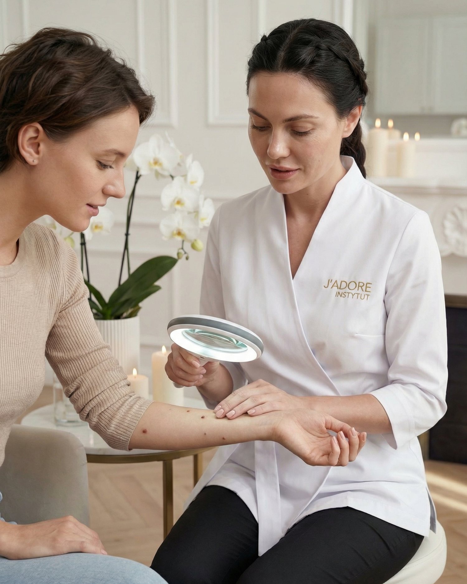
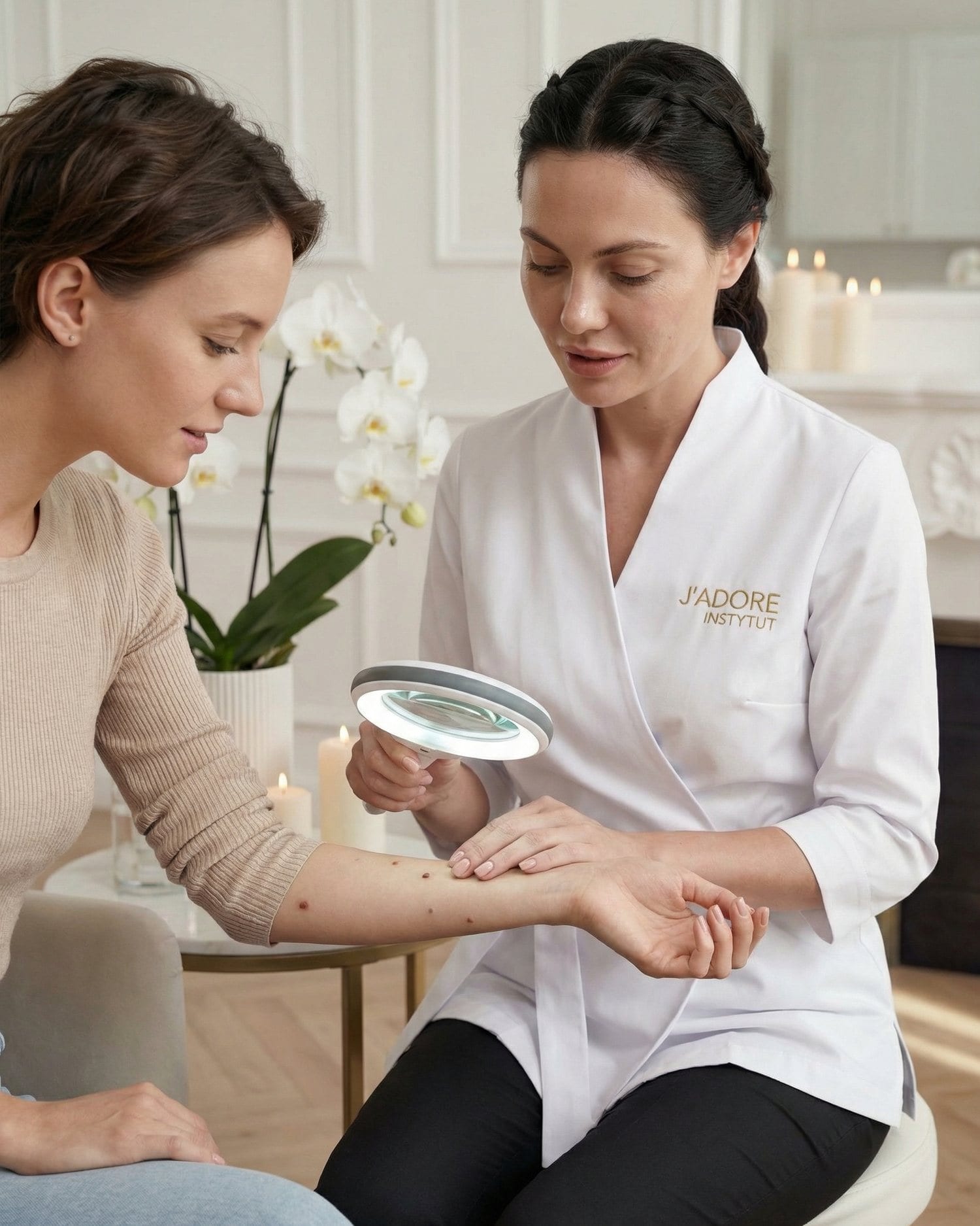
Moles, warts, fibromas, keratoses — most skin lesions we see in the salon really are benign. Over time, this builds a belief: "I know what a dangerous lesion looks like." The problem is that this very belief is one of the biggest risks. Melanocytic lesions — those made up of pigment-producing cells — can look completely innocent for a long time. A dermatoscope magnifies the image dozens of times and eliminates light reflections, revealing patterns the naked eye simply can't register.
Six myths — because these are the ones we hear most often
Myth 1: "A wart is a wart — why examine it?" This belief holds true in the vast majority of cases. Indeed, most small lesions on the neck, décolletage or face are benign fibromas or seborrheic warts. The trouble is, they look similar to early lesions that aren't benign at all. Without dermatoscopy, the difference is invisible. Removal takes a moment — but the consequences of skipping the examination can be serious and long-lasting. That's why dermatoscopy isn't a formality — it's a safety measure.
Myth 2: "The examination is only needed for large or dark lesions." Colour and size are poor risk indicators. Early lesions that need attention can be small and pale. Some melanocytic lesions show no concerning features on visual inspection for a long time — and that's exactly what makes them particularly deceptive. Dermatoscopy assesses structural patterns — pigment distribution, network symmetry, border character — not just colour or dimensions.
Myth 3: "If it doesn't hurt or itch, nothing bad is happening." The absence of subjective symptoms has no diagnostic value in the early assessment of skin lesions. Most benign lesions never cause any discomfort — but the same is true of very early lesions that need verification. Waiting for symptoms is a high-risk strategy in this context. That's why a regular mole check and dermatoscopy before any removal make sense even for people with no symptoms at all.
Myth 4: "Dermatoscopy is only needed before laser removal, not surgical removal." The removal method doesn't change the need for the examination. The decision on whether a lesion should be removed at all, and whether and where to remove it, must be preceded by a dermatoscopic assessment — regardless of the technique. What's more, with surgical removal it's possible to take a sample for histopathological analysis, which is an added safeguard — but histopathology itself doesn't replace the initial assessment and treatment planning.
Myth 5: "I had this same mole checked a year ago — the result was fine, so I can skip it now." Skin lesions evolve. A mole that looked stable a year ago may have since changed structure in ways invisible to the naked eye. Dermatoscopy assesses the current state, not the history. That's why every examination is only valid at the moment it's performed — and why, for regular mole checks, repeated dermatoscopy over time is more valuable than a single examination from years ago.
Myth 6: "A beauty salon won't do dermatoscopy properly anyway." That's an imprecise generalisation. Dermatoscopy performed in a cosmetology salon, by trained specialists with the right equipment, allows for a reliable initial assessment. What matters most: the examiner's qualifications, the quality of the equipment and — crucially — a readiness to refer you to a dermatologist wherever the result raises doubts. A good salon doesn't replace a dermatologist — it works alongside one: assessing, documenting, referring when needed.
What the examination looks like in practice
- 01
Consultation & review of lesions
A short conversation about the history of the lesions — when they appeared, whether they've changed, your risk factors (sun exposure, family history). The specialist examines all lesions, not just the ones you mention.
- 02
Dermatoscopic examination
Every lesion intended for removal or raising any doubt is examined with a dermatoscope. The specialist assesses structural patterns and documents the result — a photo or a description.
- 03
Qualification
Three possible outcomes: a benign lesion — qualified for treatment; a lesion requiring monitoring — recommendation for regular review; a lesion raising doubts — referral to a dermatologist before any intervention.
- 04
Treatment or referral
If qualification is positive, removal can take place the same day. If there's any doubt — a medical consultation comes first, and only then a decision about treatment. This isn't bureaucracy — it's safety.
When "by eye" isn't enough — two scenarios
Visual assessment
What's visible to the naked eye
- Colour and outline of the lesion
- Approximate dimensions
- Clear asymmetries
- Very advanced lesions
Dermatoscopy
Instrument-based examination under magnification
- Pigment network patterns
- Symmetry and border character
- Vascular structures
- Regression zones and globules
- Features visible only under a dermatoscope
What happens when a salon skips dermatoscopy
A salon that removes every reported lesion without an initial assessment is acting against safety standards — even if it operates smoothly and painlessly. This isn't about scaremongering: the vast majority of lesions removed are benign. But precisely because they're benign, a great many can be removed for years without a single negative event. And that builds a false sense that the examination is unnecessary.
Yet the only moment when a lesion requiring a different path from cosmetic removal can be identified is before the treatment. After laser removal, there's no material left to analyse. After surgical removal, the material can be examined histopathologically — but only if someone has arranged for it to be preserved and assessed. Dermatoscopy is the gateway: it decides what happens next — remove here, monitor, or refer to a doctor.
Dermatoscopy versus regular mole checks — different purposes, the same value
Pre-treatment dermatoscopy and a dermatoscopic mole check are two different, though related, examinations. A mole check makes sense as a periodic screening — especially for people with numerous moles, fair skin, a history of intense sun exposure, or a family history. Pre-treatment dermatoscopy is mandatory before every removal — regardless of risk factors.
- In summer — strong sun exposure can activate melanocytic lesions; a check at the start of summer and after it is good practice
- For new lesions that have appeared in recent months — dermatoscopy before any decision to remove or monitor
- For lesions that you feel have changed colour, shape or size — immediately, without waiting for your next appointment
- Before every removal — no exceptions
Dermatoscopy and other treatments near moles
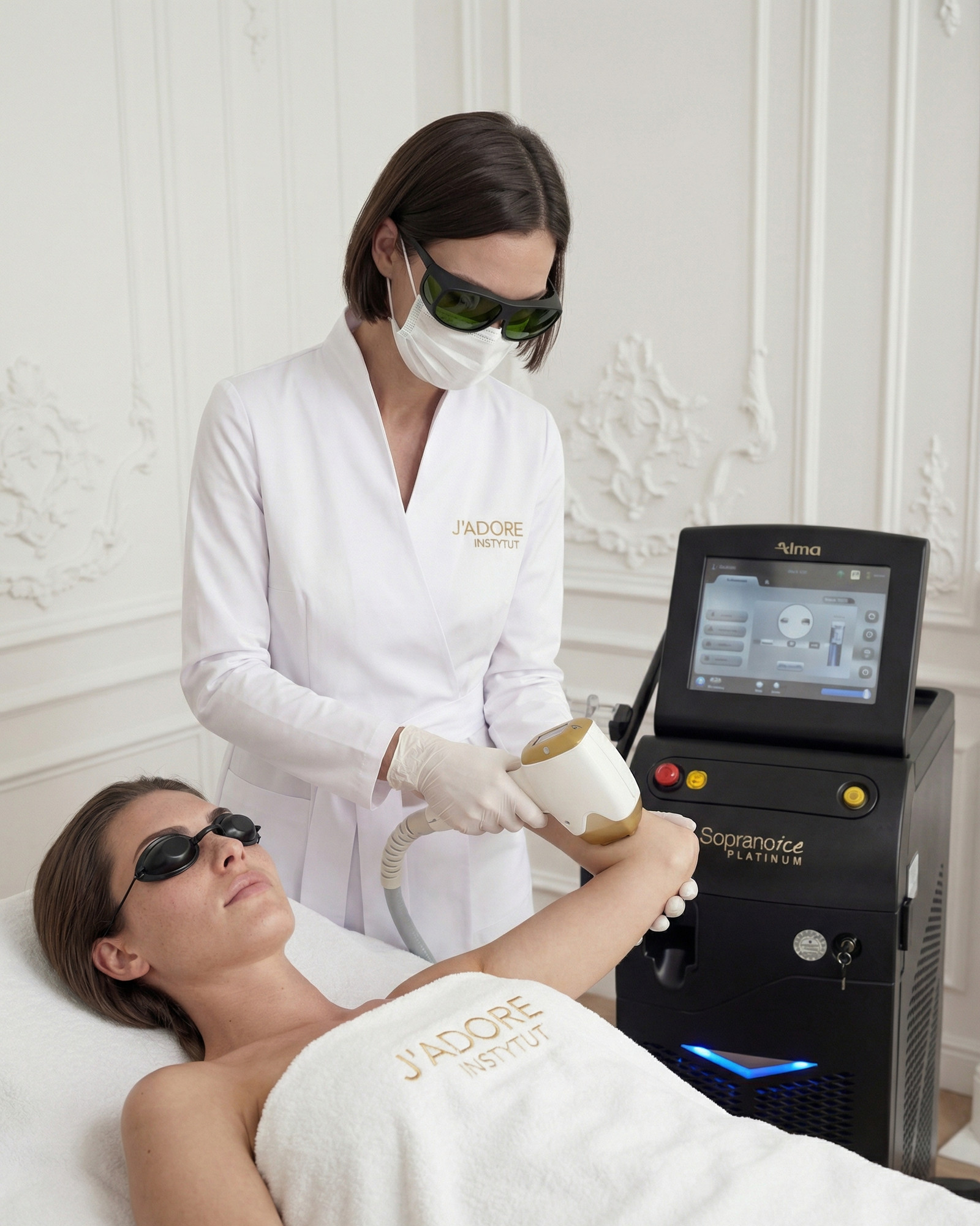
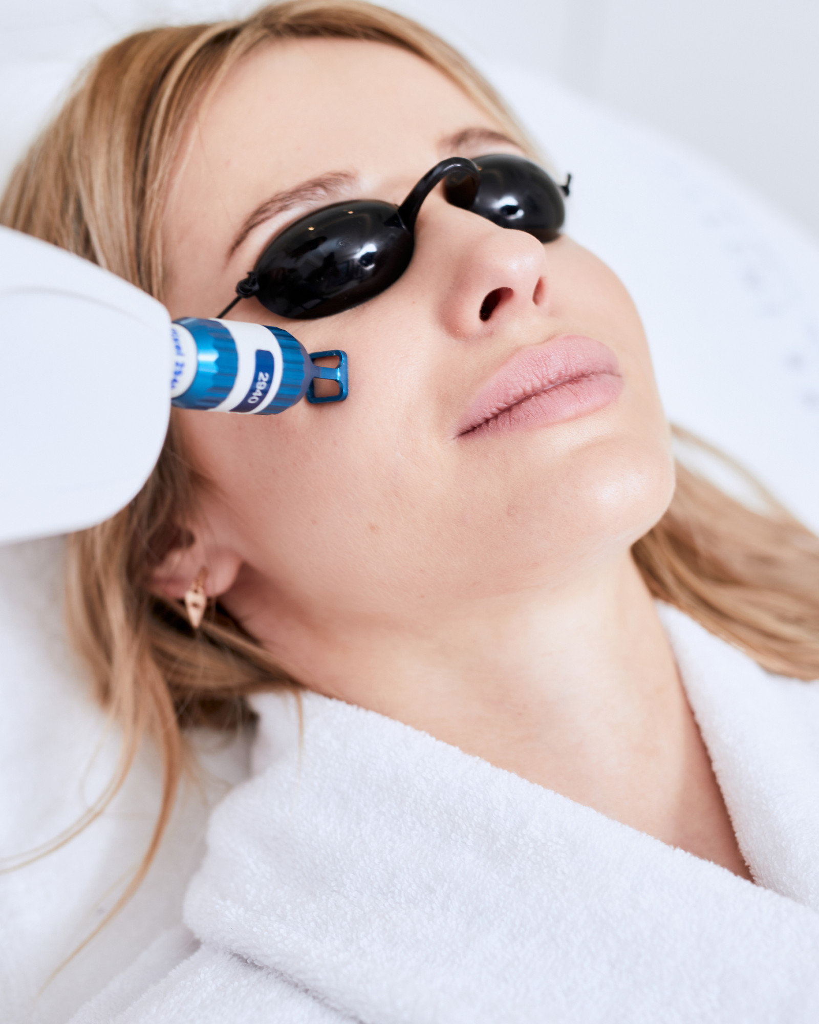
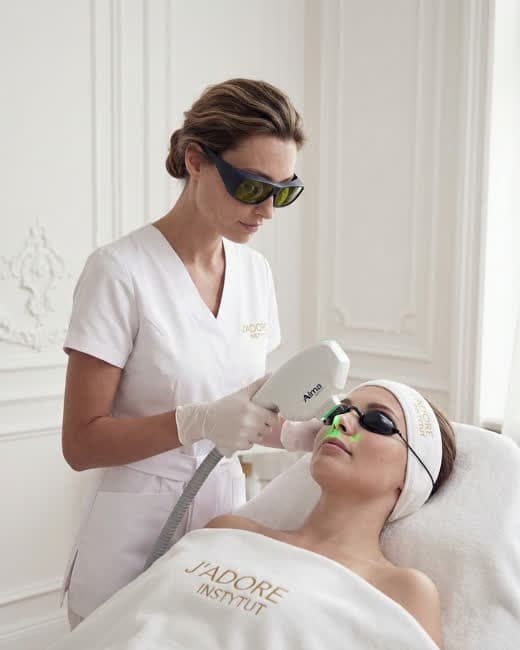
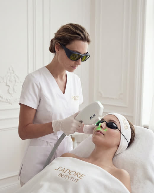
A separate, often overlooked issue is laser treatments and peels near moles. Using a laser on an area of skin that contains a mole requires a prior dermatoscopic assessment — not because the laser is dangerous for every mole, but to make sure the mole is suitable for laser therapy in that area. A professional salon always maps moles before any laser treatment on the body.
Laser removal of lesions
Dermatoscopy is a condition of qualification — without it, laser removal of a lesion shouldn't go ahead.
Laser hair removal
For laser hair removal near numerous moles, an initial dermatoscopic assessment helps determine a safe protocol and identify areas to avoid.
Chemical peels on the body
Before deeper peels on areas with numerous moles, it's worth having a check first — this is standard practice in reliable salons.